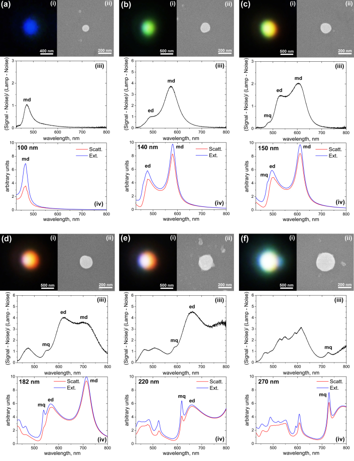
Figure 3. Close-view dark-field microscope (i) and SEM (ii) images of the single nanoparticles selected in Fig. 2.
Figures 3 (a) to (f) correspond to nanoparticles 1 to 6 from Fig. 2 respectively. (iii) Experimental dark-field scattering spectra of the nanoparticles. (iv) Theoretical scattering and extinction spectra calculated by Mie theory for spherical silicon nanoparticles of different sizes in free space. Corresponding nanoparticle sizes are defined from the SEM images (ii) and noted in each figure.

