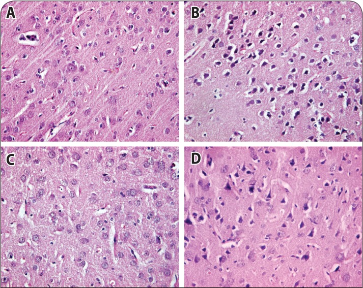
Figure 7.
Photomicrographs of brain histopathology of control, DDVP exposed and treated rats, H&E 40×. A: Control rat brain showing normal neuroglial cells arranged in several layers. B: DDVP exposed rat brain showing chromatolysis of nuclear material, gliosis and pyknotic neurons in cortex. C: Rat brain with DDVP + 2-PAMCl and atropine showing mild lesions of neuronal damage with moderate gliosis. D: 2-PAMCl + atropine + curcumin (200 mg/kg) treatment after DDVP exposure showing mild gliosis and chromatolysis along with minimal lesions of neuronal degeneration.

