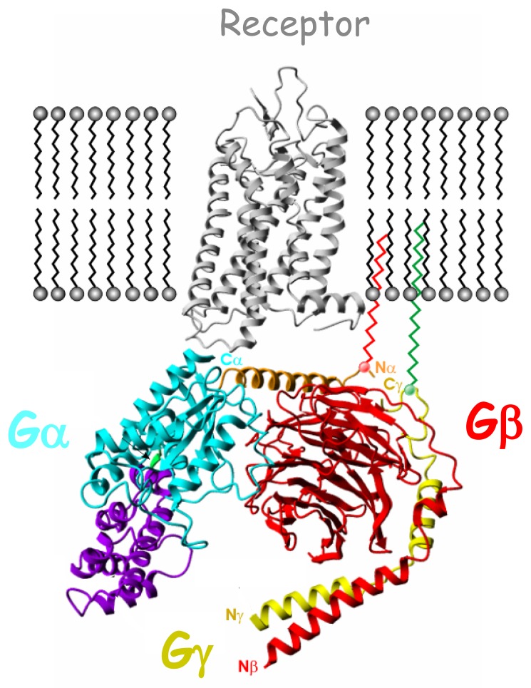
Fig. 1.
Schematic complex in the plasma membrane between rhodopsin (gray; PDB code 1GZM) and the inactive heterotrimeric G protein composed of αi1, β1, and γ2 subunits (light blue/violet, red and yellow respectively; PDB code 1GG2). Gαi1 N-terminal helix (αN) is shown in brown, while Gαi1-GTPase and Gαi1-helical domains (αi1H) are in light blue and violet respectively. Linker 1 connecting Gαi1-GTPase to the Gαi1H is represented in green. Both Gαi1N and Gγ2 C-terminal helix (γ2C) are anchored to the membrane through lipid modification.

