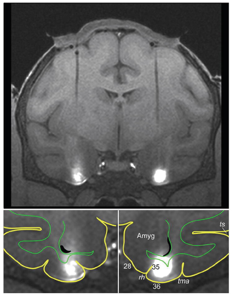
Figure 1.

MRI-guided targeting of the monkey’s perirhinal cortex. Upper panel: MR image of a coronal brain section showing (i) bilateral tracks left by guide tubes through which the injection cannulae were lowered and (ii) bilateral infusions of the MR contrast agent gadolinium (white areas). The cap-shaped object above the brain is postoperative granular-tissue growth between the dura mater and the guide-grid chamber (the latter being invisible to MR in this image taken after the gadolinium was removed from the chamber). Lower panels: Enlarged views of the ventral part of the section shown in the upper panel. Yellow lines outline the brain’s pial surface, and green lines, the border between grey and white matter. As shown in these views, the infusions were limited largely to the perirhinal cortex, located in the lateral half of the inferior temporal gyrus (i.e. the gyrus between tma and rh). Abbreviations: 28, Brodmann area (BA) 28 or entorhinal cortex; 35, BA 35 or perirhinal cortex in the lateral bank of the rhinal sulcus; 36, BA 36 or perirhinal cortex on the gyral surface; Amyg, amygdala; rh, rhinal sulcus; tma, anterior middle temporal sulcus; ts, superior temporal sulcus.
