Fig. 2.

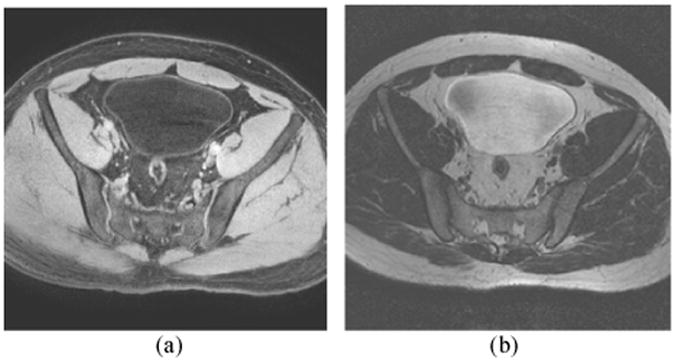
Typical bladder MR images. (a) T1-weighted MR image. (b) T2-weighted MR image. The bladder wall is more distinguishable in the T1 image than in the T2 image.

Typical bladder MR images. (a) T1-weighted MR image. (b) T2-weighted MR image. The bladder wall is more distinguishable in the T1 image than in the T2 image.