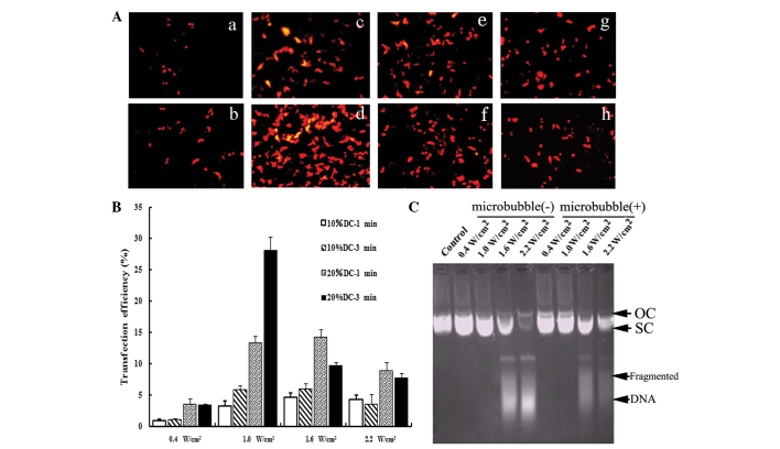
Figure 1.
Optimization of the UTMD-mediated delivery system in vitro. (A) Fluorescence microscopy views under various ultrasound intensities (0.4, 1.0, 1.6 and 2.2 W/cm2) and exposure times (1 and 3 min), DC=20%. With the optimal ultrasound parameters (Fig. 1a-d), the transfection efficiency was increased significantly (28.04±2.27%). (B) Effects of ultrasound parameters on the transfection efficiency. When cells were irradiated using 10% DC with 0.4 W/cm2, only a few expressed RFP. When the ultrasound intensity was increased to 1.0 W/cm2 or when the DC was 20%, the transfection efficiency increased significantly (P<0.05). However, when the ultrasound intensity was further increased to 1.6 and 2.2 W/cm2 or the exposure time was extended to 3 min, the expression of RFP decreased. As compared with 10% DC, irradiating for 1 and 3 min with 20% DC improved the expression rate significantly (P<0.05). (C) Effects of ultrasound parameters on the integrity of plasmid DNA. Ultrasound parameters, 1.0 W/cm2; DC=20%; exposure time, 3 min. The structures and migration rates of vectors did not markedly change following irradiation with 0.4 and 1.0 W/cm2. Following treatment with higher ultrasound intensity, the structures of the vectors were markedly destroyed and DNA fragments could be observed. Under the same ultrasound parameters, the addition of SonoVue had no effect on the integrity of plasmid DNA. UTMD, ultrasound-targeted microbubble destruction; DC, duty cycle; RFP, red fluorescence protein; OC, open circular DNA; SC, supercoiled DNA.

