
Figure 1.
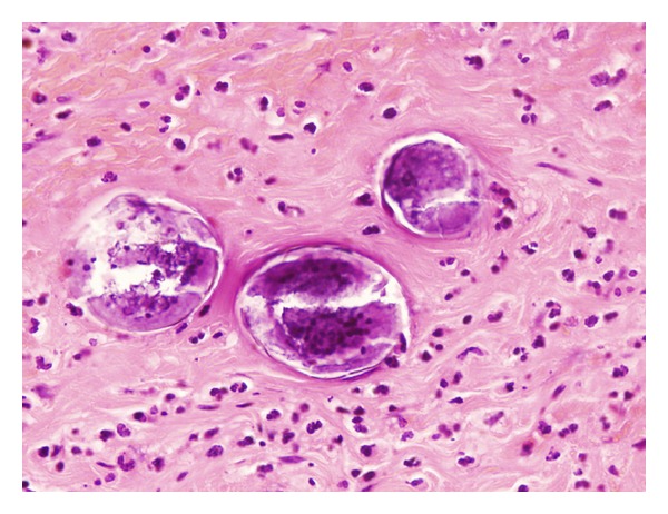
Paraffin embedded 5 μm thick section of appendiceal wall, process from the patient appendix. Magnified image by Light microscopy ×350 and stained with Haematoxylin and Eosin saffron (H-E). The identification of a lateral spine on an oval egg (at the end of an oval egg) is consistent with S. mansoni.