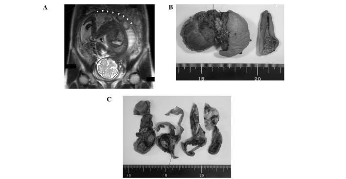
Figure 1.
Macroscopic features of the sacrococcygeal teratoma. (A) Coronal section of the fetus in the uterus using magnetic resonance imaging (MRI). The mass on the bottom of the fetus is indicated by white arrowheads. (B) Whole image of the tumor and (C) cross-sectional surface. The tumor had dark-yellow solid components and cystic lesions.

