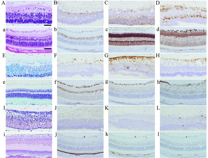
Figure 3.
Histopathological images of the retina-like structure in the human teratoma (A-L) with a normal mouse retina as a control (a-l). (A, a) H&E staining, (E, e) Kluver-Barrera staining, (I, i) silver staining. Immunohistochemistry for (B, b) Pax6, (C, c) synaptophysin, (D, d) β-tubulin, (F, f) HuC/D, (G, g) nestin, (H, h) GFAP, (J, j) RPE65, (K, k) HMB45 and (L, l) Ki-67. Scale bars in (A) 50 μm; in (a), 75 μm.

