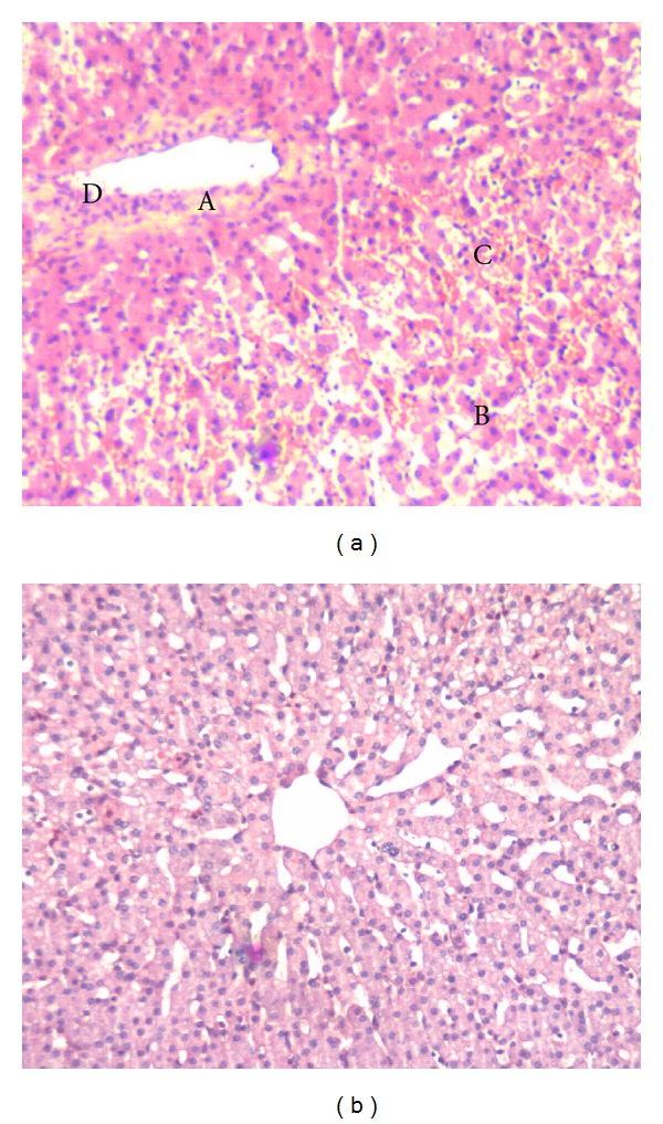
Figure 3.

Liver injury eight hours after reperfusion. Liver tissue was taken 8 hrs after reperfusion and processed for light microscopy by H&E staining. (a), control; (b), pONS; controls displayed severe focal necrosis (A) with disintegration of hepatic cords (B), hemorrhage (C), and neutrophil infiltration (D) 8 hrs after reperfusion; this effect was significantly blunted by pONS. Pictures depict typical pattern of pathology; pONS: preconditioning oral nutritional supplement.
