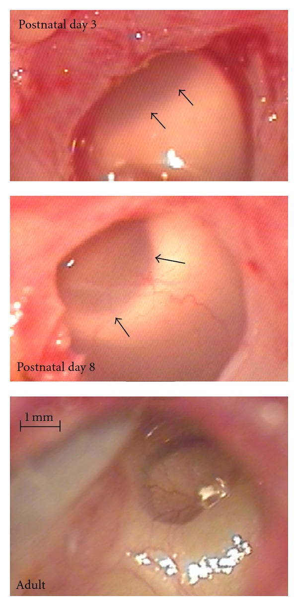
Figure 2.

Photographs of the bulla in animals of the youngest group, compared to an adult animal. At postnatal days 0 and 3 (top), the bulla was filled with milky viscous tissue that covered the round window (its rims show through, marked by arrows). At postnatal day 8 (middle), the tissue was more translucent. In all older animals investigated, the bulla was fully pneumatized and did not show any further developmental changes (bottom).
