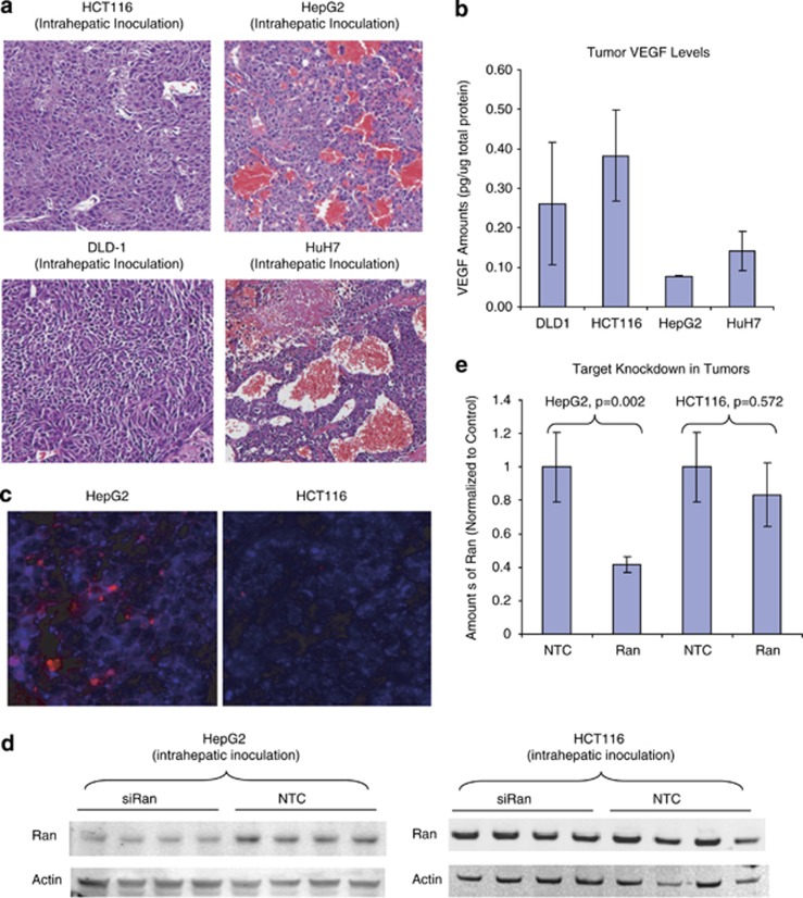
Figure 3.
SNALP triggers better target knockdown in highly vascularized tumors. (a) Liver tumors were established via intrahepatic injection of HCT116 or HepG2 cells in scid mice. Representative hematoxylin and eosin sections of the HCT116- and HepG2-derived liver tumors were shown. (b) VEGF levels in tumor homogenates (N=5 for each tumor type) were determined using the QuantiGlo human VEGF kit and normalized to the total protein amount in the same sample. (c) SNALP liposome containing DY647-labeled TetR siRNA were administrated to tumor-bearing mice (intravenous (i.v.), 2.5 mg siRNA kg−1). Tumors were harvested 20 h later and cryosections of the tumor were stained with an antibody that recognizes a human specific mitochondria marker. Representative confocal image of resulted HepG2 or HCT116 sections were shown. Blue: human mitochondria staining that specifically labels xenograft tumor tissue. Red: DY647 signal that indicates the presence of SNALP(siRNA) delivery. (d) Mice that bear HCT116- and HepG2-derived liver tumors were administrated with SNALPs-containing NTC or siRAN at 2 mg siRNA kg−1, i.v., q.d. × 2. Tumors were collected 48 h after last dosing and analyzed by western blotting for the expression of Ran and Actin. N=4 for each treatment group. (e) Densitometry quantification of western results in (d). The y axis represents the average amount of Ran (normalized to the amount of actin in each sample).

