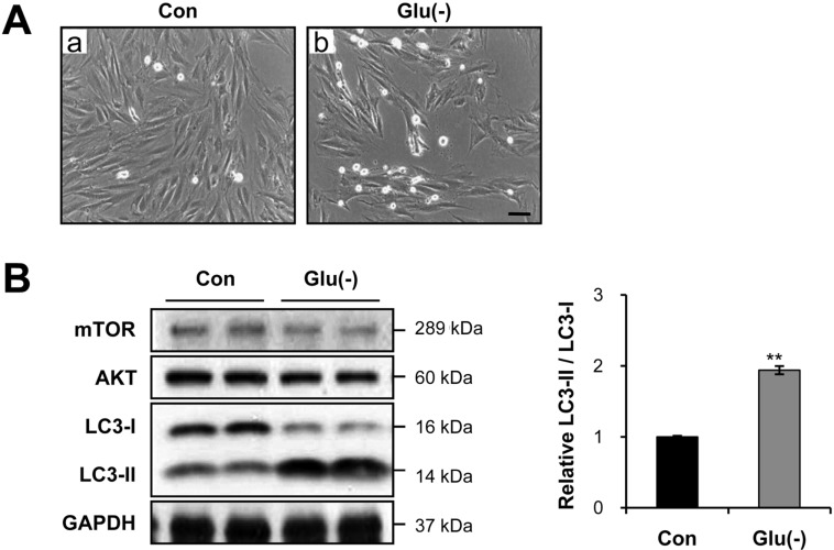
Figure 1.
Glucose starvation strongly induces cell-size reduction and autophagy. A, Representative inverted microscopy images after 12 h of glucose starvation. Magnification ×100. Scale bar=40 µm. B, Immunoblot analysis of microtubule-associated protein-1 light chain-3 (LC3)-I and LC3-II. Cardiac myocytes were incubated with complete media (control) or treated with glucose-free [Glu(-)] medium for 12 h. In A and B, the results represent data from three independent experiments. **P<0.01

