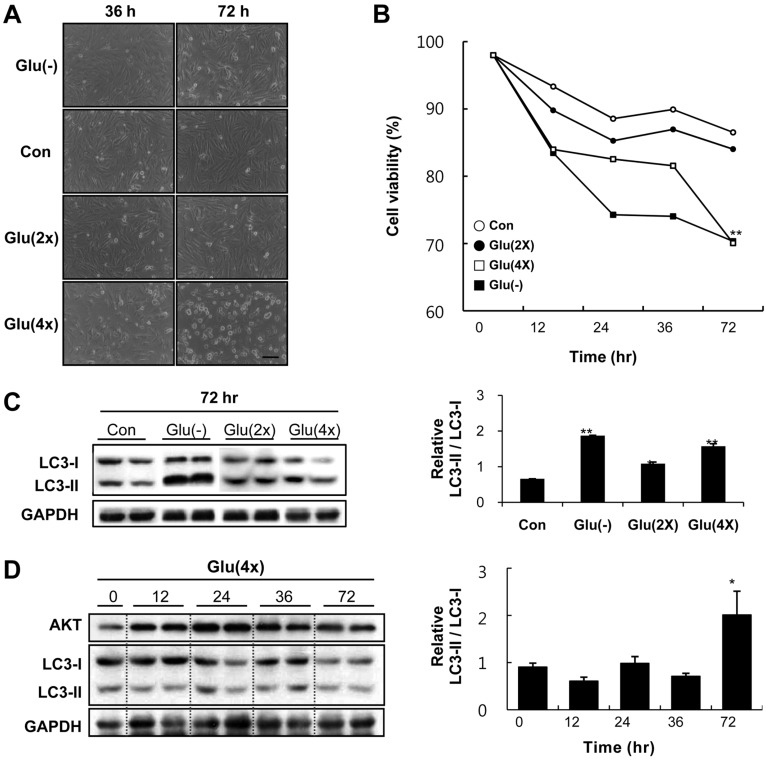
Figure 2.
High glucose media suppresses proliferation of cardiomyocytes, and induces cell death with accompanied by autophagy. A, Representative light microscopy images of H9c2 cells. Cells were treated with complete media as a control (Con) or with glucose-free [Glu(-)], 33 mM glucose [Glu(2×)], or 66 mM glucose [Glu(4×)] media for 36 hr and 72 hr. Scale bar = 100 um. B, Time course of cell viability after media exchange, estimated by trypan blue assay. Values are mean from three independent experiments. **P<0.01 vs. Con. C, Immunoblot analysis of LC3-I and LC3-II. Cells were cultured in complete media as a control (Con), glucose-free media [Glu(-)], 33 mM glucose [Glu(2×)], or 66 mM glucose [Glu(4×)] for 72 hr, and quantitative analysis of relative LC3-II expression to compare the level of autophagy between groups. *P<0.05 vs. Con, **P<0.01 vs. Con. D, Time course of LC3 expression by immunoblot analysis after 66 mM glucose [Glu(4×)] treatment, and quantitative analysis of relative LC3-II expression to compare the level of autophagy between time points in Glu(4×) group. *P<0.05 vs. Con

