Figure 1A.

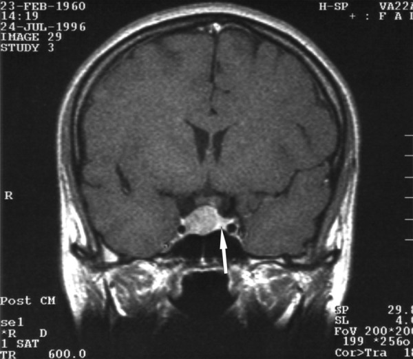
A T1-weighted image after constrast medium administration, coronal plane: the adenoma is visible, compressing and shifting the pituitary gland to the left side. The arrow indicates the compressed pituitary.

A T1-weighted image after constrast medium administration, coronal plane: the adenoma is visible, compressing and shifting the pituitary gland to the left side. The arrow indicates the compressed pituitary.