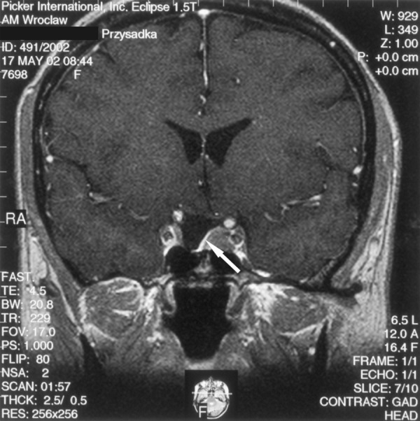
Figure 2.

Condition post pituitary adenoma surgery performed 5 years before. A T1-weighted image after contrast medium administration, coronal plane: a narrow band of the gland connected to the hypothalamic infundibulum is visible. The hypothalamic infundibulum is slightly shifted to the right. On the left side of the sella there is a hypointensive structure visible, suggesting a residual tumour. The arrow points at a spared, small fragment of the pituitary gland. However, lack of progression in 7-year-long MRI follow-up allowed diagnosis of post-surgical fibrosis.
