Figure 4B.

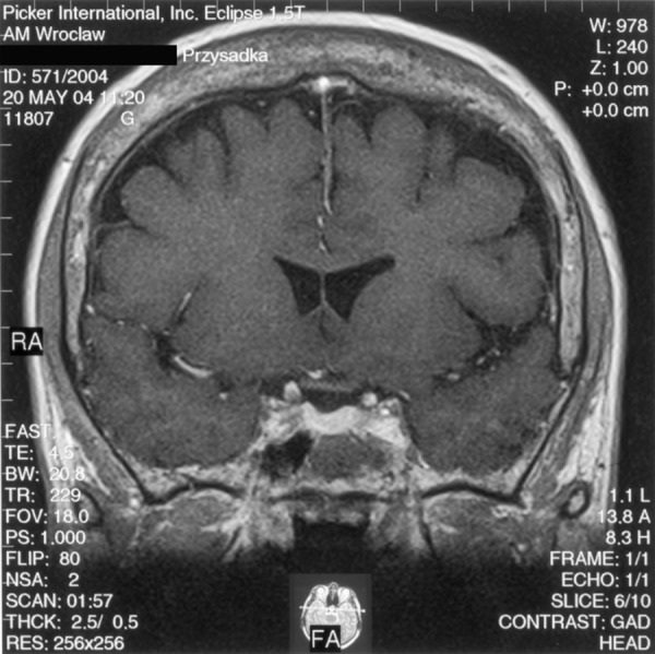
The same patient – condition approx. 1.5 years after the pituitary tumour resection. A T1-weighted image after contrast medium administration, coronal plane: normal pituitary gland visible, uniformly enhanced.

The same patient – condition approx. 1.5 years after the pituitary tumour resection. A T1-weighted image after contrast medium administration, coronal plane: normal pituitary gland visible, uniformly enhanced.