
Figure 5.
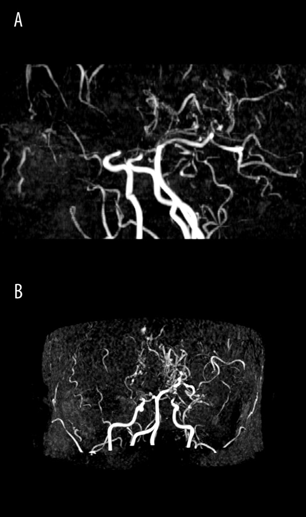
Case 1, Suzuki grades III/IV. MR angiography, ToF (time-of-flight) technique, MIP reconstructions in sagittal (A) and axial plane (B). No signal from either of the anterior cerebral and middle cerebral arteries. Collateral vessels of “moyamoya” type, visible at the base of the brain.