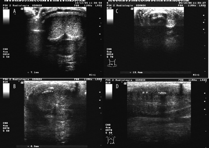
Figure 1.
Ultrasound image of the penis in a patient with Peyronie’s disease. (A) a plaque of type 1 – a typical thickening of the tunica albuginea without acoustic shadowing. (B) a plaque of type 2 – a moderately calcified plaque with slight acoustic shadowing. (C) a plaque of type 1 – a severely calcified plaque with typical shadowing. (D) 2 plaques – one of type 2 and one of type 3.

