Figure 2. .
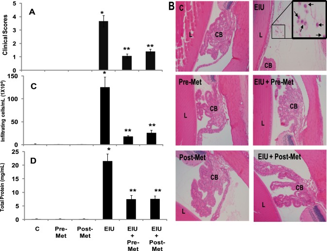
Metformin prevents LPS-induced infiltration of inflammatory cells and increase in protein levels in AqH. (A) The pathologic score of EIU in Lewis rat eyes injected with LPS in the absence and presence of metformin was determined at 24 hours with a slit lamp microscope. Results are given as mean ± SD (n = 6). #P < 0.001 versus control. **P < 0.001 versus EIU (Wilcoxon–Mann-Whitney test). (B) Histopathological results of paraffin-embedded sections showing infiltrated cells (inset) in the anterior chamber of EIU rat eyes without or with metformin injected 12 hours before or 2 hours after LPS administration. H&E-stained serial sections of rat eyes were photographed under a light microscope. Magnification, ×200. (C) The infiltrated inflammatory cells were determined by trypan blue exclusion cell counting and (D) total protein levels in the AqH. Results are expressed as the mean ± SD (n = 5); *P < 0.001 versus the control group; **P < 0.05 versus the EIU group. C, control; Pre-Met, pretreatment with metformin; Post-Met, posttreatment with metformin; EIU, endotoxin-induced uveitis; EIU + Pre-Met, endotoxin-induced uveitis + pretreatment with metformin; EIU + Post-Met, endotoxin-induced uveitis + post treatment with metformin; CB, ciliary body; L, lens.

