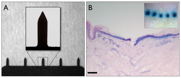
Figure 1. Microneedle morphology and skin penetrative capabilities.
(A) Bright field micrographs of an in-plane row of five microneedles, each with a height of approximately 750μm, insert shows in greater detail the microneedle geometry. (B) En face (insert) and 10μm H&E stained histological section of human skin treated with microneedles and post-stained with methylene blue (bar = 300μm).

