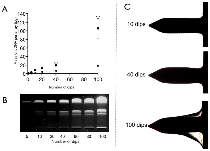
Figure 3. Microneedles dip-coated coated with pDNA.
(A) UV-spectrophotometric quantification of pDNA dip-coated onto microneedles with increasing number of dips and varying drying time from 5 seconds (open circles) to 30 seconds (closed circles). Data presented as mean ± SD (n=4); One way analysis of variance, ***P < 0.01. (B) Gel electrophoresis of pDNA recovered from microneedles following surface coating with varying number of dips (30 seconds drying time). (C) Bright field micrographs showing the build up of pDNA on the surface of the microneedles when the number of dips was increased (30 seconds drying time).

