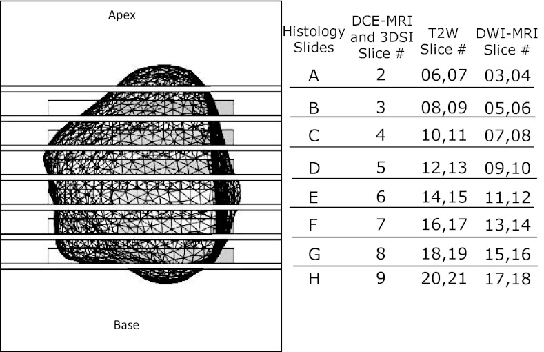
Figure 2.
Top view of the patient-specific mold showing the prostate cavity (wire mesh), the slots for the knife every 6 mm, and correspondence of the locations of 5 mm histology slices to the locations of MP-MRI slices. The rectangular outlines near each slot indicate the location of optional windows that were also visible in the MRI slices shown in Figs. 3d, 3e, 3f, 4d, 4e, 4f.

