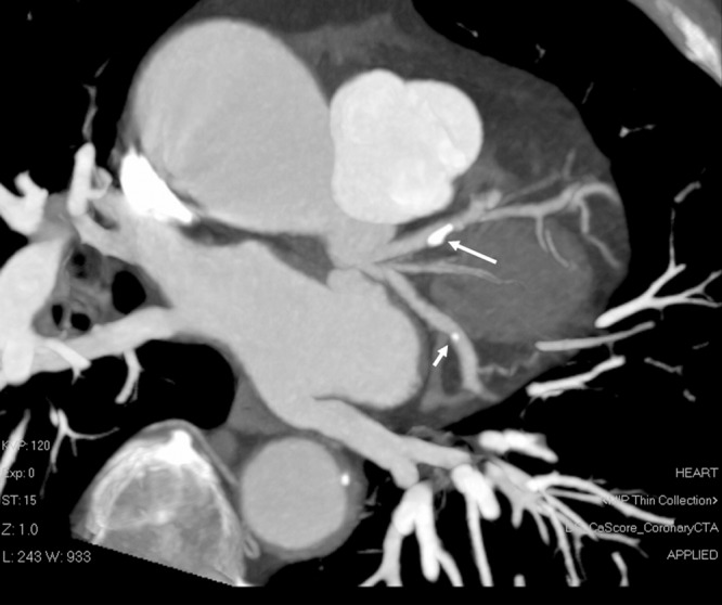
Figure 2. Calcified coronary plaques are shown at the proximal segment of left anterior descending (long arrow) and distal segment of left circumflex (short arrow) branches on a coronal maximum-intensity projection image in a 73-year old man with chest pain and history of hypertension.

