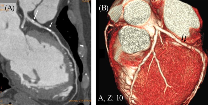
Figure 3. A mixed coronary plaque (long arrow in A) is present within a lesion at the proximal segment of left anterior descending artery as shown on a curved planar reformatted image in a 55-year-old man with symptoms of chest discomfort and epigastric pain. A calcified plaque is also noticed at the mid-segment of the same left coronary artery (short arrow in A). Three-dimensional (3D) volume rendering demonstrates significant stenosis in the left anterior descending due to the calcified plaque (arrows in B).

