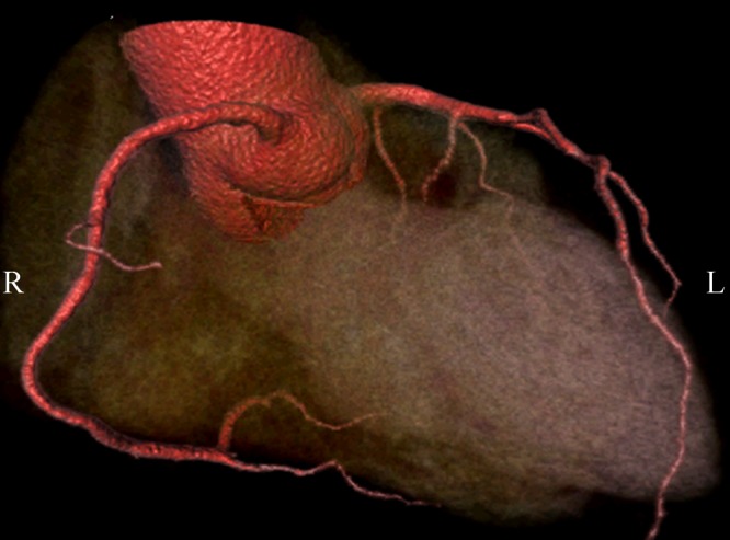
Figure 6. 3D volume rendering of the coronary arteries and side branches are clearly demonstrated with use of 320-slice CT angiography in a 58-year-old man presenting with chest pain. Volumetric data are acquired within a single heart beat with excellent image quality.

