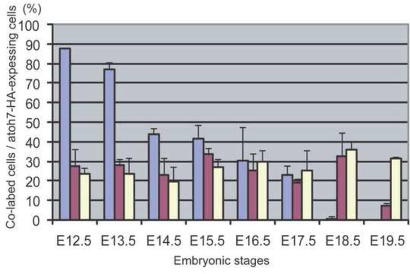
Figure 3.
Histogram showing the fraction of Atoh7 -expressing RPCs in the neuroblast layer of the developing retinas that are co-expressing Pax6 (blue-gray), Chx10 (red) or Neurod1 at the indicated developmental times. Anti-HA, anti-Pax6, and anti-Neurod1 antibodies were used to label the retinas (n=3) and the fraction of cells expressing the indicated transcription factor was determined as described in the Methods section.

