
Fig. 1. Schematics and photographs of multisomes.
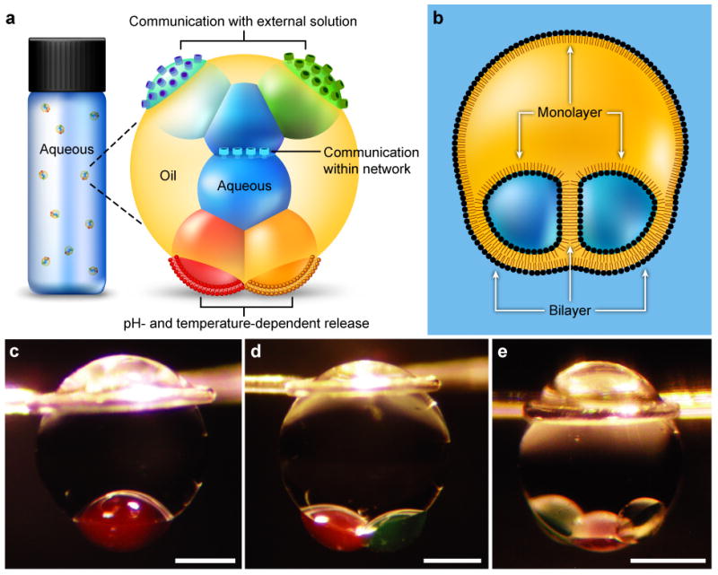
a, Illustration of a multisome. Aqueous droplets encapsulated in an oil drop are connected by lipid bilayers, which allow the droplets in the network to communicate through protein pores. Pores in bilayers at the surfaces of the aqueous droplets that protrude from the oil drop enable the network to communicate with the bulk solution. Multisomes can release the contents of encapsulated droplets by pH- or temperature-induced rupture of bilayers. b, Schematic of an encapsulated two-droplet network, illustrating the lipid monolayers and bilayers. c–e, Photographs of multisomes containing one (c), two (d) and three (e) inner droplets. Oil drops were suspended on wire loops to allow extended study. Aqueous droplets were dyed with 25 μM sulphorhodamine 101 (red) or fluorescein (green). Scale bars = 400 μm.