
Fig. 4. Communication by diffusion through αHL pores.
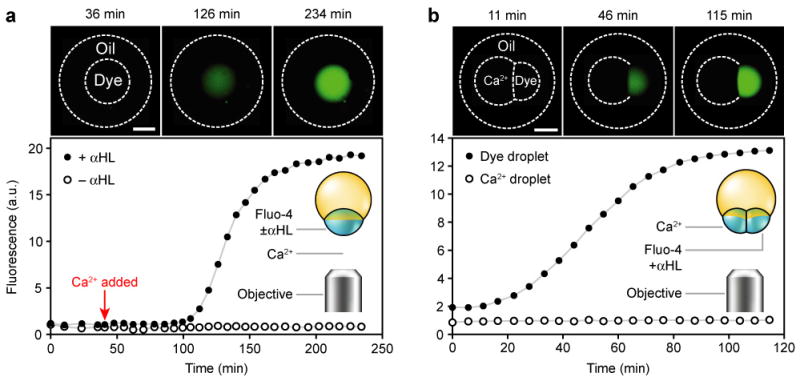
Fluorescence photographs and measurements of multisomes. Oil and inner droplets are outlined in the photographs where they are invisible. Inner droplets containing dextran-conjugated fluo-4 or Ca2+ are respectively labelled ‘Dye’ or ‘Ca2+’. a, Two multisomes with a single inner droplet each, in the same bulk solution; both multisomes contained the dye, and one also contained αHL. The photographs are of the droplet containing αHL, while the graph includes measurements from both droplets. Following the addition of Ca2+ to the external solution, the droplet containing αHL increased in fluorescence over ~1.5 h, while the droplet without protein did not. Scale bar = 300 μm. b, A multisome containing a two-droplet network, in which one droplet contained Ca2+ and the other contained the dye and αHL. The dye-containing droplet increased in fluorescence, while Ca2+-containing droplet did not. Scale bar = 300 μm.