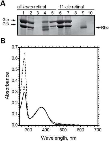
Figure 5.
The dependence of complex formation on activation of the N2C,E113Q,D282C mutant. (A) SDS-PAGE analysis of fractions from the purification of the rhodopsin N2C,E113Q,D282C mutant on 1D4-Sepharose in the presence of transducin, as visualized by Coomassie blue staining. The rhodopsin mutant was reconstituted with either all-trans-retinal (lanes 1–5) or 11-cis-retinal (lanes 5–10), as is indicated in the figure. Lanes 1 and 6, transducin sample that was applied to the 1D4-matrix; lanes 2 and 7, unbound material; lanes 3 and 8, last wash; lanes 4 and 9, 1D4-peptide eluate; lanes 5 and 10, GTPγS eluate. Only the α- and β-subunits of transducin are shown. (B) UV/Visible absorption spectra for the purified rhodopsin fractions. Spectrum 1, N2C,E113Q,D282C mutant reconstituted with all-tran-retinal (corresponding to fraction in A, lane 4); spectrum 2, N2C,E113Q,D282C mutant reconstituted with 11-cis-retinal and purified in the dark (corresponding to fraction in A, lane 9).

