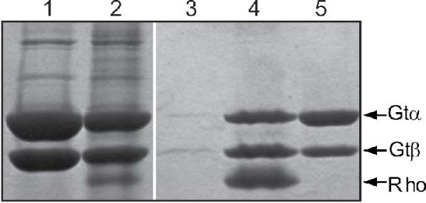
Figure 9.

Purification of the activated rhodopsin/transducin complex in 2.5% DMPC/DHPC bicelles. The figure shows SDS-PAGE analysis of fractions from 1D4 immunoaffinity purification of the N2C,E113Q,D282C rhodopsin/transducin complex stained with Coomassie blue. The protocol was as described in Experimental Procedures using 2.5% DMPC/DHPC bicelles. Lane 1, transducin fraction applied to the column; lane 2, unbound material; lane 3, last wash before elution of the complex; lane 4, 1D4-peptide eluate; lane 5, GTPγS eluate. Only the γ- and β-subunits of transducin are shown. It should be noted here that in control experiments using buffers without bicelles no protein could be eluted from the immunoaffinity matrix with the 1D4-peptide.
