Figure 2.

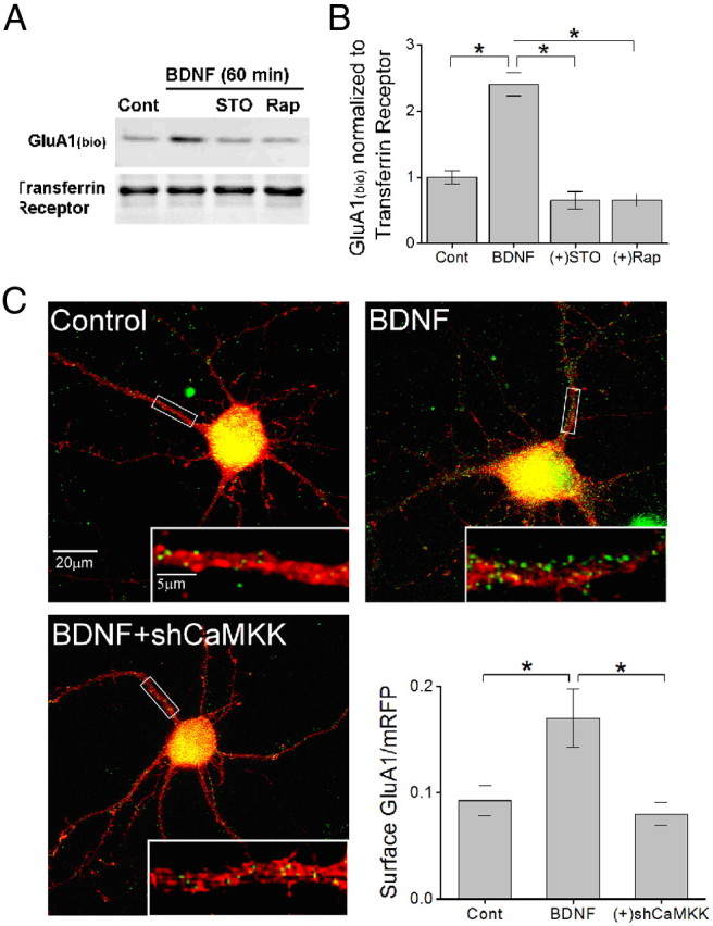
CaMKK mediates BDNF-induced increases in GluA1 surface expression. A, Representative immunoblot of biotinylated surface GluA1 (GluA1bio) and surface transferrin receptor following 60 min of BDNF treatment in the absence or presence of STO-609 or Rap as shown. B, Quantification of surface biotinylated GluA1 normalized to surface transferrin receptor for indicated conditions (n = 5 from 5 independent experiments). C, Representative immunofluorescence images of hippocampal primary neurons (10 DIV) transfected with monomeric RFP (red) and superimposed with surface GluA1 (pseudo-colored in green) for control. Neurons treated with 50 ng/ml BDNF plus or minus coexpression of short hairpins to α and β CaMKK (shCaMKK). Inset shows a magnification of the boxed segment of proximal dendrite. Lower right, Group data for surface GluA1 levels for indicated conditions (n = 8–12 neurons per coverslip from 3 independent experiments). Error bars indicate SEM *p < 0.05 by Student's t test.
