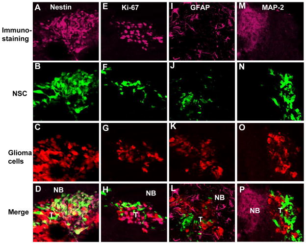
Fig. 6. Engineered hNSC do not differentiate in mouse glioma model.
hNSC-aaTSP-1 or control hNSC-GFP-Rluc were implanted in the close vicinity of established Gli36-EGFRvIII-FD gliomas. Representative images of brain sections of hNSC-aaTSP-1 mice sacrificed on day 12 and immunostained with nestin, Ki67, GFAP and MAP-2 antibodies. Different panels showing the expression of tumor cells (red), hNSC (green) and nestin, Ki67, GFAP or MAP-2 immunostaining (purple). NB-normal brain; T-tumor.

