Fig. 2.
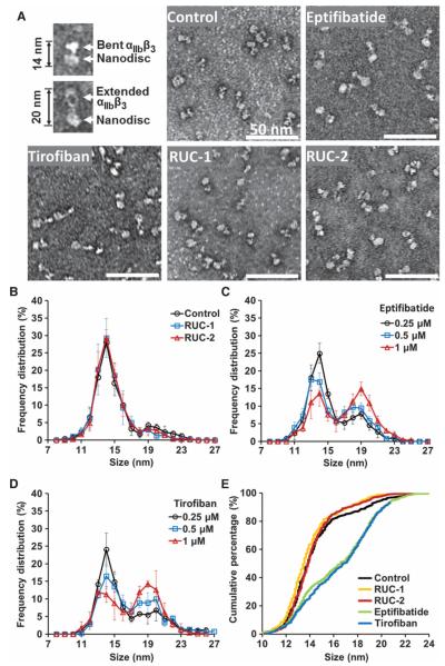
Negative stain EM of αIIbβ3 nanodiscs in the absence and presence of αIIbβ3 antagonists. (A) Representative images of bent and extended αIIbβ3 nanodiscs and images of αIIbβ3 nanodiscs in the presence of buffer, eptifibatide (1 μM), tirofiban (1 μM), RUC-1 (100 μM), or RUC-2 (10 μM). (B to D) Quantitative measurements of αIIbβ3 NIL values in the absence and presence of αIIbβ3 antagonists. (B) NIL value distributions in the presence of buffer, 100 μM RUC-1, or 10 μM RUC-2. (C and D) Dosedependent NIL value distributions in the presence of eptifibatide (C) or tirofiban (D). The mean ± SD of five separate experiments is depicted for each condition; a total of 600 to 700 particles contained in five separate electron microscopic images were measured at × 33,000 magnification in each experiment. (E) Cumulative percentage of NIL values in the presence of buffer, 100 μMRUC-1, 10 μM RUC-2, 1 μM eptifibatide, or 1 μM tirofiban.

