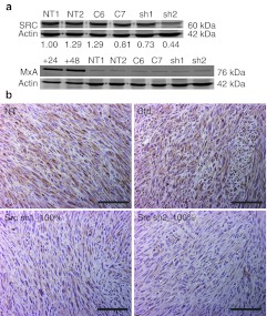
Figure 3.
Expression of Src and interferon-responsive MxA in mouse tumors. (a) Western blot was used to analyze Src and MxA protein expression from tumor lysates. β-Actin was used as a loading control. Normalization of Src by actin is shown below each lane of the corresponding blot. Lanes: NT1-2 = nontransduced tumors, C6−C7 = control virus transduced tumors, sh1 = tumor transduced with shRNA1 against Src, sh2 = tumor transduced with shRNA2 against Src, +24 and +48 = interferon-induced cell lysates collected at 24 and 48 hours postinduction (positive controls). (b) Immunostaining against Src, 200× magnification, scale = 100 µm. Ctrl, transduced with a control vector expressing shRNA against luciferase; NT, nontransduced; sh1, shRNA sequence 1 against Src; sh2, shRNA sequence 2 against Src; shRNA, small hairpin RNA.

