
Figure 5.
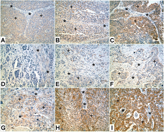
Immunoperoxidase staining of ZIP8 in high grade urothelial carcinoma.A &B. Noninvasive urothelial carcinoma showing weak staining of ZIP8 (+). Asterisks (*) indicate stromal tissue which is negative for ZIP8. C. Noninvasive urothelial carcinoma with focal moderate staining of ZIP8 (+). Asterisks (*) indicate stromal tissue which is negative for ZIP8. D. Invasive urothelial carcinoma with negative staining of ZIP8 (−). Few smooth muscle fibers between the invasive tumor nests are weakly positive for ZIP8 (*). E. Invasive urothelial carcinoma with focal weak staining of ZIP8 (+). Asterisks (*) indicate stromal tissue which is negative for ZIP8. F. Invasive urothelial carcinoma with weak staining of ZIP8 (+). A few spindled shaped stromal cells are also weakly positive for ZIP8 (*). G. Anaplastic urothelial carcinoma showing moderate staining for ZIP8 (+). Asterisks (*) indicate stromal tissue which is negative for ZIP8. H. Invasive urothelial carcinoma with moderate to strong staining of ZIP8 (*). A few spindled shaped stromal cells are weakly positive for ZIP8 (*). I. Poorly differentiated urothelial carcinoma with strong staining for ZIP8 (+). Asterisks (*) indicate stromal tissue which is negative for ZIP8.