
Figure 7.
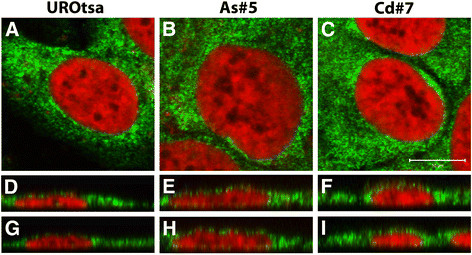
Immunofluorescent localization of ZIP8 in UROtsa parent and transformed cell lines. ZIP8 was found to concentrate heavily in the paranuclear region of the cells along with a punctate staining that extended throughout the cytoplasm, indicative of association with the endoplasmic reticulum. The parent UROtsa cells are shown in A,D,G while the transformed cells As#5 are shown in B, E, H and Cd#7 are shown in C, F, I. ZIP8 staining is shown in green while the nuclear dye To-PRO-3 iodide is shown in red. Single Z-series slices are shown in A - C while orthogonal views in the X- and Y-planes are shown in D-F and G-I respectively. Bar = 10 μm and represents the scale for A - C.