Figure 1.
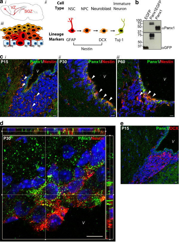
Panx1 is expressed in nestin-positive periventricular NSC/NPCs throughout postnatal development in vivo. (A) Mouse brain schematic with the neurogenic ventricular zone (VZ) and subgranular zone (SGZ) regions highlighted (i). Cell types throughout neurogenesis and the lineage markers expressed at each stage. Relatively quiescent NSCs become highly proliferative NPCs, and neuronally committed migratory neuroblasts before exiting the cell cycle as immature neurons (ii). Schematic of cell types in the periventricular region. The ependymal cell layer is interdigitated by thin processes or whole luminal surfaces of the radial glia-like NSCs (iii). This figure is a partial reproduction of another figure created by the same authors that appears in [2] and is reproduced with permission. (B) Western blot of lysates from HEK293T cells overexpressing EGFP, Panx1EGFP, or Panx1, showing specificity of the Panx1 Cterm antibody. (C) Confocal images of murine brain slices at the periventricular region. Nestin-positive NSCs exhibit high levels of Panx1 co-expression in postnatal day 15 (P15) (i), P30 (ii) and P60 (iii) brains. Arrows indicate areas of co-expression. (D) Image from a confocal z-stack with orthogonal side-views of a P30 periventricular region. Panx1 expression is noted in ependymal/sub-ependymal layers. (E) Confocal image from a P15 periventricular region. Panx1 is absent from DCX-positive neuroblasts. Hoechst 33342 was used as a nuclear counterstain in C-E. All scalebars 10 μm. DCX, doublecortin; NPC, neural progenitor cells; NSC, neural stem cells; Panx1, pannexin 1; V, ventricle.

