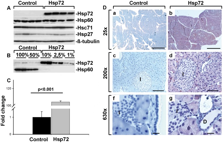
Figure 1. Hsp72 mice display robust Hsp72 overexpression in pancreatic acinar cells.
A) Pancreatic tissue homogenates from control and Hsp72 mice were analyzed using antibodies against Hsp72, Hsc71, Hsp27, Hsp60 and ß-tubulin representing a loading control). B) Serial dilutions of pancreatic tissue homogenates were made to estimate the level of Hsp72 overexpression using two independent control and Hsp72 animals. Hsp60 was used as a loading control. C) Quantitative RT-PCR analysis of pancreatic RNA extracts confirmed the strong Hsp72 overexpression in Hsp72 mice. 4 mice per group were used and the values were normalized to the Hsp72 levels in control mice, which were arbitrarily set as 1. D) Representative tissue sections of pancreata from nontransgenic (left column) and Hsp72 mouse (right column) were stained with antibody against Hsp72. Note the robust Hsp72 overexpression in pancreatic acinar cells, but not islets (I in c,d,f,g) or ductal cells (D in g) of Hsp72 animals. Scale bars 1 mm (a,b), 100 µm (c,d) and 200 µm (f,g).

