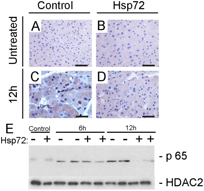
Figure 9. Hsp72 mice display attenuated NF-κB signalling.
Immunohistochemical staining (A) depicts nuclear localization of p65 which serves as a marker of NF-κB activation. Nontransgenic (left column) and Hsp72 mice (right column) were analyzed prior to (upper row) and 12 hours after caerulein administration (lower row). Scale bar 50 µm. (B) To further quantify the extent of NF-κB activation, pancreatic nuclear extracts were incubated with p65 antibody. Note the attenuated NF-κB signalling in caerulein-treated Hsp72 mice when compared with their non-transgenic littermates. HDAC2 was used as a loading control.

