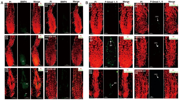
Figure 6. HFs of the estrogen treated mice showed activation of BMP pathway.
(A) Increase expression of BMP4 in hair keratinocytes and DP cells after 3 and 4 days of estrogen treatment vs. HFs of the oil treatment mice. (B) After 3 and 4 days of estrogen treatment, a high phosphorylation level of BMP signaling effector, Smad 1/5, was detected in precortex, while the control ones had negligible P-Smad 1/5 expression in ORS. The days of treatment are indicated. Propidium iodide-labeled nuclei are in red. ORS: outer root sheath; IRS: inner root sheath; E2: 17β-estradiol; HF: hair follicle; DP: dermal papilla. Scale bar: 50 µm (A); 20 µm (B).

