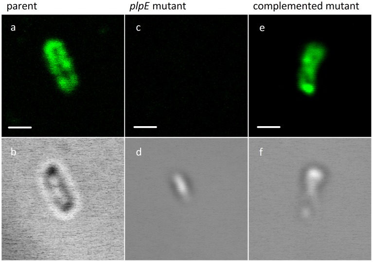
Figure 2. Surface localisation of P. multocida PlpE by immunofluorescence assay.
Bacteria were fixed with paraformaldehyde, incubated with chicken antiserum against PlpE, stained with Alexa Fluor 488 goat anti-chicken IgG, and visualized by fluorescence microscopy. Fluorescence (panels: a, c, e) and DIC (Differential Interference Contrast) (panels: b, d, f) selected images of the bacterial Z-stack cross-sections. Control staining with antiserum raised against an unrelated protein showed no surface fluorescence (data not shown). Scale bar = 1 µm.

