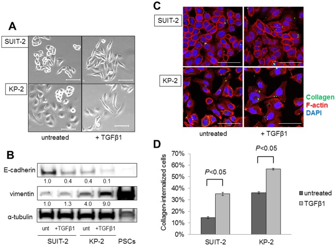
Figure 2. Treatment with TGF-β1 induces EMT in SUIT-2 and KP-2 cells and enhances their collagen internalization.
(A) SUIT-2 and KP-2 cells are transformed to a spindle-shaped morphology following treatment with TGF-β1 for 72 h. Scale bars: 100 µm. Original magnification: ×200. (B) Western blotting analysis showing that E-cadherin expression is reduced and vimentin expression is increased in SUIT-2 and KP-2 cells by TGF-β1 treatment. The fold differences in the representative immunoblots quantified by densitometry are shown below the respective lanes. unt: untreated. (C) SUIT-2 and KP-2 cells were cultured with or without TGF-β1 for 72 h, followed by incubation with OG-gelatin (green) for 2 h. The cells were fixed and stained with Alexa Fluor 647-conjugated phalloidin (red) to visualize the cell outlines. Fluorescence microscopy reveals that the collagen internalized by SUIT-2 and KP-2 cells is increased after treatment with TGF-β1. Scale bars: 100 µm. Original magnification: ×400. (D) Quantitative data for collagen internalization by SUIT-2 and KP-2 cells determined by flow cytometry reveals significant differences in the collagen uptake ability between untreated and TGF-β1-treated cancer cells. Each cell line was analyzed in triplicate and the percentages of collagen-internalized cells are expressed as means ± SD. Comparisons between untreated and TGF-β1-treated cells were carried out using Student's t-test. All experiments were performed more than three times and representative images are shown.

