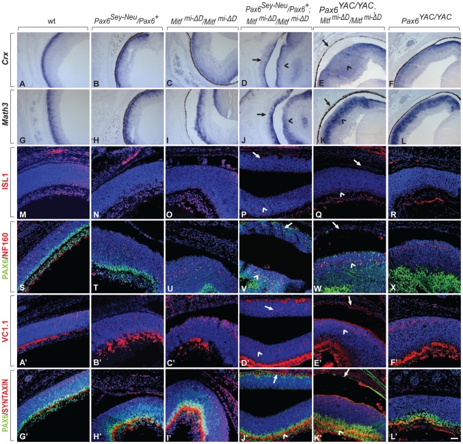
Figure 4. Development of a differentiated laminated retina in Pax6Sey-Neu/Pax6+;Mitfmi-ΔD/Mitf mi-ΔD but not Pax6YAC/YAC;Mitf mi-ΔD/Mitf mi-ΔD mice.
(A–L) Sections of eyes from P0 mice of the indicated genotypes were subjected to in situ hybridization for Crx, a photoreceptor marker (A–F) or Math3, an amacrine cell marker (G–L). Note that the ectopic staining is not present in the RPE of Pax6YAC/YAC;Mitf mi-ΔD/Mitf mi-ΔD mutants (compare arrows in D,J with E,K for ectopic staining; arrowheads mark normal retinal staining). (M–L′) Immunofluorescent labeling for the indicated markers on P0 eye sections of the indicated genotypes. ISL1 is a ganglion cell marker (M–R), as is PAX6 at this time point (S–X, G′–L′, green). NF160 marks horizontal cells (S–X, red); VC1.1 marks amacrine cells (A′–F′, red); and SYNTAXIN marks synapses (G′–L′, red). Arrows mark the transdifferentiating portions of the RPE in Pax6Sey-Neu/Pax6+;Mitf mi-ΔD/Mitf mi-ΔD mice (P,V,D′,J′) or the corresponding non-transdifferentiating portions in Pax6YAC/YAC;Mitf mi-ΔD/Mitf mi-ΔD mice. The normal retinas continue to express each of these markers (arrowheads in the corresponding figures). Scale bar (A–L): 115 µm; (M–X, A′–L′): 90 µm.

