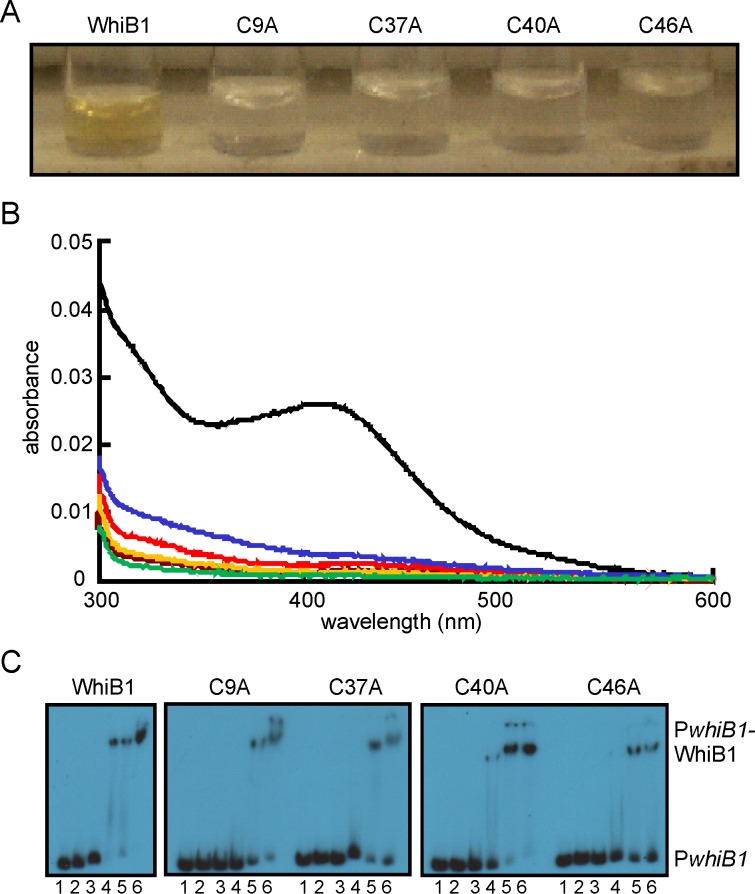
Figure 1. All four Cys residues of WhiB1 are required for iron-sulfur cluster acquisition.
(A) Photograph depicting anaerobic reconstitution of WhiB1 and variants with the indicated single Cys substitutions after removal of unincorporated components by chromatography on heparin Sepharose. (B) UV-visible spectra of WhiB1 and variants after anaerobic reconstitution of iron-sulfur clusters. The black line shows the spectrum of wild-type WhiB1 (4.5 µM); the blue line, WhiB1-C9A (2.5 µM); the red line, WhiB1-C37A (3.7 µM); the brown line, WhiB1-C40A (3.6 µM); the orange line, WhiB1-C46A (3.6 µM). The green line shows the spectrum of apo-WhiB1 (2.3 µM). The buffer was 25 mM Tris-HCl pH 7.4 containing 0.5 M NaCl and 10% glycerol. (C) All four WhiB1 Cys variants bind whiB1 promoter DNA (PwhiB1). Radiolabeled PwhiB1 DNA was incubated with increasing concentrations of the indicated WhiB1 proteins before separation of protein-DNA complexes in electrophoretic mobility shift assays. Lanes 1, no protein; lanes 2–6 contain, 1, 2, 5, 10 and 15 μM WhiB1, respectively. The locations of PwhiB1 and PwhiB1-WhiB1 complexes are indicated.

