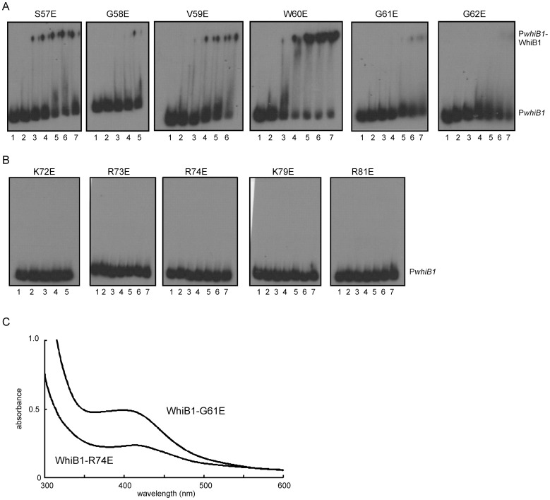
Figure 3. DNA-binding and iron-sulfur cluster acquisition by WhiB1 variants with amino acid substitutions in the C-terminal region.
WhiB1 proteins with the indicated amino acid substitutions were incubated with radiolabeled PwhiB1 DNA and complexes were separated by electrophoresis. (A) Amino acid substitutions in the putative β-turn of WhiB1. (B) Amino acid substitutions in two conserved amino acid motifs located downstream of the predicted β-turn of WhiB1. Lanes 1, no protein; lanes 2–7, 2.5, 5, 7.5, 10, 12.5 and 15 μM of the indicated WhiB1 protein, respectively. The locations of PwhiB1 and PwhiB1-WhiB1 complexes are indicated. (C) Representative UV-visible spectra of holo- WhiB1-G61E (26 μM; A420∶A280 ratio 0.19) and WhiB1-R74E (13 μM; A420∶A280 ratio 0.16).

