
Figure 4. FRET-detected monomer association of PFO and PFOR468A.
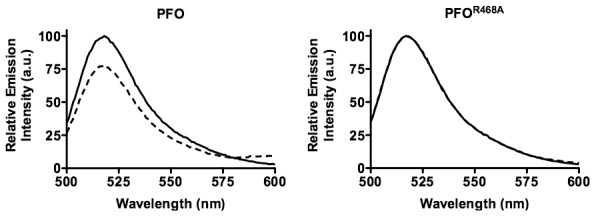
A cysteine was substituted for Asp-30, located in domain 1 and the derivatives were labeled with Alexa Fluor 488 (donor, D) or Alexa Fluor 568 (acceptor, A).A 4∶1 molar ratio of A-labeled PFOR468A (dashed line) or unlabeled PFOR468A (U; solid line) to D-labeled toxin was incubated in the presence of human erythrocyte ghost membranes and fluorescence emission intensity of D was measured.