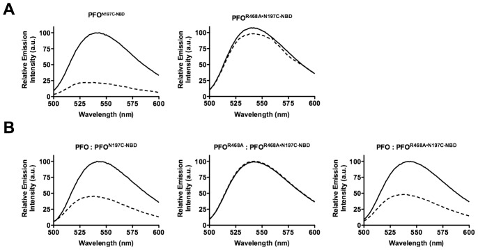
Figure 5. Disruption of the D2/D3 interface in PFO and PFOR468A.
(A) A cysteine was substituted for TMH1 residue Asn-197, which is located within the D2/D3 interface. Each derivative was labeled with NBD and incubated in the presence (dashed line) and absence (solid line) of human erythrocyte ghost membranes. If TMH1 breaks its contact with D2 then Asn-197 moves from a buried, nonpolar location at the interface with D2 to the lumen of the pore. An NBD positioned at this location will therefore undergo a nonpolar to polar transition, which results in the quenching of the fluorescence emission. (B) Unlabeled native PFO or PFOR468A were mixed at a 4∶1 molar ratio with PFON197C-NBD and PFOR468A•N197C-NBD derivatives. The fluorescence emission intensity of NBD was measured from 500 to 600 nm. These data are representative of 3 experiments.

