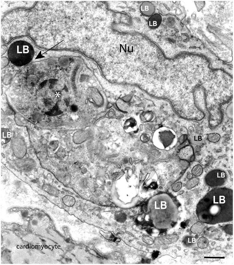
Figure 3. Lipid bodies (LBs) increase in number and interact with phagosomes within heart inflammatory macrophages during parasite infection.
LBs with different sizes are seen as electron-dense or electron-lucent organelles surrounding and in contact (arrow) with a large phagolysosome containing an intact amastigote (*), the intracellular form of the parasite Trypanosoma cruzi. Rats were infected with the Y strain of T. cruzi and samples of the heart, a target organ of the parasite, processed for transmission electron microscopy at day 12 of infection [6], [68], [69]. Nu, nucleus. Scale bar, 800 nm.

