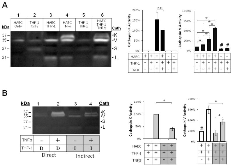
Fig 1.
TNFα and direct monocyte adhesion induced cathepsin K and V activities in endothelial cell-monocytes co-cultures. Endothelial cells, THP-1 monocytes, and co-cultures were conditioned with 10ng/mL TNFα. Monocytes were allowed to interact either (A) directly (indicated by “D”), or (B) indirectly, suspended above in a Transwell insert with a 0.2μm pore size (indicated by “I”). (A) Cell lysates were collected and loaded for cathepsin zymography. Cathepsin K active enzyme bands were quantified with densitometry and normalized to HAEC, THP-1, TNFα samples, and cathepsin V active enzyme bands were normalized to unstimulated endothelial cell controls (n=7, *p<0.05, # represents significant difference from EC control, SEM bars shown). (B) Lysates from Transwell cultures were also collected and loaded for zymography and active enzyme quantified with densitometry (n=3, *p<0.05, SEM bars shown).

